
X-rays and other imaging technologies are critical tools in the diagnosis of various medical conditions. From common issues like toothaches to complex diseases such as breast cancer, the use of X-rays has revolutionized the way physicians identify and treat conditions. Without these imaging technologies, physicians and patients alike would be left to speculate and rely on guesswork to make informed medical decisions. Thanks to X-rays, patients can receive accurate diagnoses and appropriate treatments, leading to better health outcomes.
Facts About X-rays
 Physicians are able to utilize various types of X-rays to see inside your body without resorting to an incision, and the benefits of properly administered diagnostic images far outweigh any risks. They normally are performed in a hospital radiology department, a dentist’s office, or a clinic that specializes in diagnostic procedures.
Depending on the type of X-ray being performed, you may be asked to drink a liquid or have a contrast dye injected. You may also need to fast and limit liquids or take medications to clear out the bowels if that is the area being examined.
What Are The Types of X-rays?
- Plain radiography, or plain x-ray
- Computed tomography, known as CT scanning
- Fluoroscopy — which produces moving images of an organ
- Mammography — an x-ray of the breasts
- Angiography — an x-ray of the blood vessels
When Is An X-ray Done?
X-rays are a crucial tool for doctors to diagnose and treat a wide range of diseases and injuries. Here are some common reasons why doctors use x-rays:
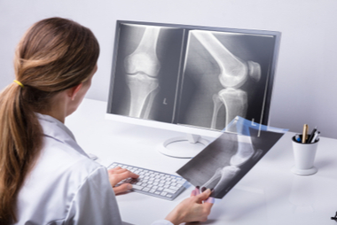
Bone Conditions
X-rays can detect fractures, dislocations, bone infections, and arthritis. They are also useful for assessing bone density and conditions like osteoporosis.
Lung Conditions
X-rays help identify lung issues such as pneumonia, collapsed lungs, and lung cancer.
Congestive Heart Failure
X-rays are used to diagnose and monitor congestive heart failure, a condition where the heart cannot pump blood effectively.
Blood Vessel Problems
X-rays can detect abnormalities like an aortic aneurysm, which is a bulge in the main blood vessel leading from the heart.
Cancer Detection
X-rays are valuable in diagnosing various cancers, including lung cancer, bone cancer, and breast cancer.
Bowel Blockages
X-rays can reveal blockages in the bowel, aiding in diagnosis and treatment.
Tooth Decay
X-rays help dentists identify tooth decay and determine appropriate dental treatments.
Foreign Object Detection
X-rays are crucial for locating and removing foreign objects, especially when a child accidentally swallows an item.
Post-surgical Checks
X-rays verify the proper positioning of wires, leads, and tubes after surgical procedures.
Conventional Radiology
This type of traditional X-ray produces a single 2-dimensional image, which you probably have seen in an actual doctor’s office if not on a medical TV show or movie. Conventional radiology is primarily used for viewing bones, bone fractures, tissues dense in calcium, dental X-rays, and the chest.
Because they are on conventional film, the doctor and patient must wait for the results. Today’s digital images (like those produced by our digital cameras) are processed by a computer, so they can be viewed immediately. As a result, they are slowing replacing film.
CT Computerized Tomography
Computerized tomography combines a traditional X-ray with computer processing to create a better resolution. A CT scan creates a series of cross-sectional images or slices to form a 3D image. This allows Southwest Diagnostic Imaging to view different parts of the body from different angles.
A CT scan can show organs, the skeleton, tissues, and any abnormalities within these systems. This type of image can show tumors and lesions in the abdomen. A CT scan may also be used to look at the following:
- The heart to discover heart disease
- The head to find an injury, blood clots leading to a stroke, tumors, or a hemorrhage
- The lungs to discover a PE or pulmonary embolism (clots), fluid, tumors, pneumonia, or emphysema
- Bones to find fractures, bone tumors, or eroded joints
Angiography
Angiography is a diagnostic medical imaging technique that involves the use of X-rays and a contrast agent to visualize the blood vessels of the body, including arteries, veins, and organs. The primary purpose of angiography is to identify and diagnose any blockages or other problems within the blood vessels that may be causing symptoms or putting the patient at risk for serious health problems.
Some benefits of an angiograph:
- Accurate diagnosis: Angiography provides a detailed and accurate picture of the blood vessels, allowing doctors to detect any blockages or abnormalities that may be affecting blood flow.
- Minimally invasive: Angiography is a minimally invasive procedure, meaning it does not require large incisions or general anesthesia, and most patients can go home the same day.
- Precise treatment planning: Once the doctor has identified the location and severity of any blockages or abnormalities, they can develop a precise treatment plan tailored to the patient’s specific needs.
- Safe and effective: Angiography is a safe and effective procedure with a low risk of complications.
- Versatile: Angiography can be used to evaluate a variety of medical conditions, including heart disease, stroke, peripheral artery disease, and aneurysms, among others.
The goal of this test is to find blockages or narrowing of blood vessels near the heart, brain, abdomen, or legs. If a blockage is discovered, SWDIC can treat it while the angiogram is being performed.
Mammography
Most women are familiar with this special X-ray known as a mammogram. It creates detailed images of the breast to be used both as a screening tool to detect cancer at an early stage, or diagnose breast disease from symptoms like pain, a lump, or discharge from the nipple.
Tumors will show up as irregular shaped white masses. Today’s mammography has advanced to include 3D images that show the entire breast. This new technology helps to increase the likelihood of early detection of breast cancers.
Fluoroscopy
Fluoroscopy produces real time images of continuous movement within the body shown on a fluorescent screen and recorded for analysis at a later time. It shows a live image of a patient’s internal structures, and can follow the path of an injected contrast substance. A fluoroscopy test depicts real time images of a beating heart or the blood flow to the muscles of the heart.
It is also used to position a pacemaker or catheter, orthopedic implants during a surgical procedure, or to view contrast agents. This is also the same technology used during a barium enema to view movement through the gastrointestinal tract.
These 5 types of X-rays are a valuable diagnostic tool for physicians that help catch diseases faster and help patients live longer! Consult with Southwest Diagnostic Imaging if you have questions about an upcoming X-ray.