
Mammography in Dallas, TX
Breast cancer is one of the most common cancers diagnosed among women in Texas. When a woman reaches the age of 40, she should have routine mammograms to evaluate her breast health. In an effort to diagnose women as soon as possible, our radiologists at Southwest Diagnostic Imaging Center uses digital and 3D mammography along with computer aided detection to detect breast cancer in its earliest stages. Our radiology team uses state-of-the-art technology to provide women in Dallas with the highest quality of breast imaging.
What is a mammogram?
Mammography (also known as a mammogram procedure) is an examination of the breast using low dose X-rays. Most medical experts agree that successful treatment of breast cancer is often linked to early diagnosis, and mammography is considered the most effective tool for early breast tumor detection. It plays a central role in the early detection of breast cancer, as it can show changes in the breast up to two years before a patient or physician can feel them.
Why Do Women Get Mammograms?
Mammograms are specialized X-ray images of breast tissue designed to detect cancer and other alterations. They serve two primary purposes:
Screening Mammogram: This type of mammogram is employed to identify potential cancerous changes in breast tissue, especially in individuals displaying no signs or symptoms. The aim is to detect cancer at an early, less invasive stage.
Determining the appropriate age to begin regular screening mammograms and their frequency is a subject of debate among experts and medical organizations. It is advisable to have a discussion with your healthcare provider regarding your individual risk factors, personal preferences, and the advantages and drawbacks of screening. Together, you can formulate a suitable screening mammography plan.
Diagnostic Mammogram: Diagnostic mammograms are conducted to investigate suspicious alterations in breast tissue, such as the discovery of a new lump, breast discomfort, unusual skin changes, nipple thickening, or nipple discharge. They are also utilized to evaluate unexpected findings identified during a screening mammogram. Diagnostic mammograms entail additional X-ray images for a more comprehensive assessment.
These examinations play a critical role in early cancer detection and ensuring appropriate follow-up care based on individual circumstances.
Offering Digital Mammography and 3D Mammography
Southwest Diagnostic Imaging Center has the capability to perform both full-field digital mammography and 3D Tomosynthesis mammography.
National Institute of Biomedical Imaging has provided very helpful information on their blog, they covered the most common questions asked by women going through this process.
Digital mammography, or 2D mammography, uses detectors that convert X-rays into electrical signals. These electrical signals produce images of the breast that can be seen on a computer screen. The radiologist is able to change the magnification, orientation, brightness, and contrast of these images.
3D mammography, or Tomosynthesis mammography, is an advanced form of breast imaging where images are acquired in 1 mm “slices” which are then reconstructed into a 3D image. The acquisition of many images in different angles provides more information per mammogram for radiologist interpretation. The positioning, compression, and radiation dose are equivalent to that of a traditional 2D mammogram.
Computer-aided detection, or CAD, uses a digitized mammographic image to search for abnormal areas of density, mass, or calcification that may indicate the presence of cancer. The CAD system highlights these areas on the images, alerting radiologists to the need for further analysis.
Frequently Asked Mammography Questions
Current guidelines from the U.S. Department of Health and Human Services (HHS), American Medical Association (AMA), and the American College of Radiology (ACR) recommend a mammography screening every year for women, beginning at age 40.
The National Cancer Institute (NCI) recommends that women who have had breast cancer and those who are at increased risk due to a genetic history of breast cancer should seek expert medical advice about frequency of screening and whether they should begin screening before age 40.
Before scheduling a mammogram, you should discuss problems with your breasts with your doctor. Generally, the best time to schedule your exam is one week following your period. Do not schedule your mammogram for the week before your period if your breasts are usually tender during this time. Always inform your mammography technologist if there is any possibility that you are pregnant.
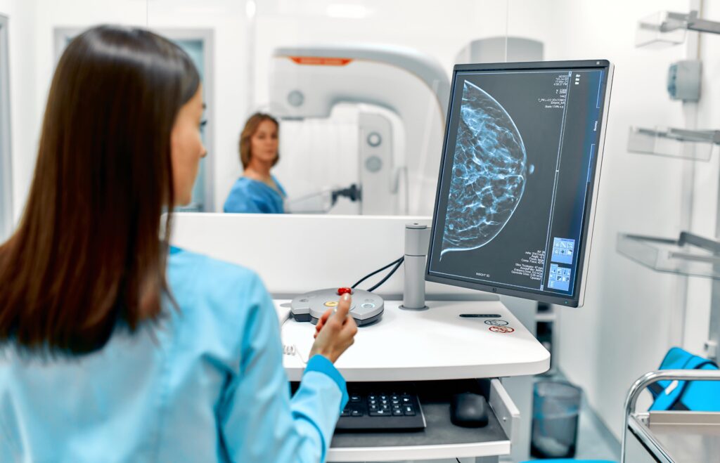
A licensed mammography technologist will perform your exam. Your breast will be placed on a special platform on the mammography unit and compressed with a clear plastic paddle. The technologist will gradually compress your breast. Breast compression is necessary in order to:
- Even out the breast thickness so that all of the tissue can be visualized.
- Spread out the tissue so that small abnormalities will not be obscured.
- Allow use of a lower X-ray dose.
- Hold the breast still to eliminate blurring of the image caused by motion.
- Reduce X-ray scatter to increase picture sharpness.
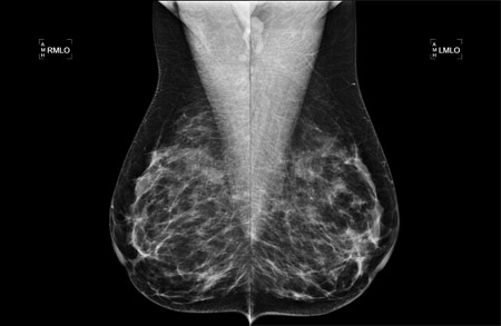
You will be positioned for two projections of each breast. Standard views are top to bottom and an oblique, or side view. Learn how you can prepare for your mammogram.
The exam takes about fifteen minutes to half an hour, depending on the number of images needed. You will feel pressure on the breast as it is squeezed by the compression paddle. Some women with sensitive breasts may experience some minor discomfort. Be sure to inform the technologist if pain occurs as compression is increased. If discomfort is significant, less compression will be used.

Schedule Your Mammogram at Southwest Diagnostic Imaging in Dallas
Southwest Diagnostic Imaging Center is a full-range, experienced, state-of-the-art imaging center in Dallas. Our mission is to provide unsurpassed patient care, the most advanced diagnostic imaging technology, and the highest standards of quality. If you are in need of a mammogram, please contact our office at (214) 345-6905 to schedule an appointment today!
Mammography Scan Hours
Mammography (Diagnostic)
Monday – Friday / 9:30 a.m. – 3:15 p.m.
Mammography (Screening Only)
Monday, Thursday, and Friday / 6:30 a.m. – 4:15 p.m.
Tuesday and Wednesday / 6:30 a.m. – 7:30 p.m.
Saturday / 9:00 a.m. – 2:00 p.m.
Schedule a Mammogram in Dallas, TX
Take an important step towards ensuring your breast health. Don’t wait, prioritize your well-being and contact our imaging clinic in Dallas, TX by calling (214) 345-6905 today!