
Whether you are having a mammogram or an MRI to detect cancer, there is someone behind the screen looking at the images and making a professional diagnosis. It’s not the Wizard of Oz, but a doctor and well-trained diagnostic radiologist who interprets the images, makes a diagnosis, and issues a report. Here’s what you should know about diagnostic radiology and cancer:.
Southwest Diagnostic Imaging Center Radiologists Behind the Screen
Radiologists are medical doctors who specialize in the diagnosis and treatment of injuries and diseases using medical imaging procedures like x-rays, computed tomography (CT), magnetic resonance imaging (MRI), nuclear medicine, positron emission tomography (PET), and ultrasound.
A radiologist uses these advanced imaging tools to find the cancer, discover if it has spread, and then monitor how your body is responding to current treatments.
Radiologists complete at least 13 years of training. They are certified by the American Board of Radiology and have specific requirements for continuing education throughout their career.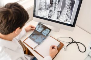
Frequently Used Imaging Tools
Southwest Diagnostic Imaging Center uses the following imaging tools at our office in Dallas to diagnose, manage, and assist your oncology care team in monitoring your progress.
CT Scans
A CT scan is one of the imaging tools used frequently by radiologists to provide information about a cancer diagnosis. CAT scans are one of the most important tools for doctors and patients. The images are used to create 3D images of the inside of your body.
Not only can the radiologist use a CT scan to diagnose cancer, but in addition:
- It can help indicate the exact place for a biopsy.
- It will provide the stage of the cancer.
- It will give an evaluation of how the treatment is progressing.
- It will check for recurrence.
Bone Scans
A bone scan will tell Southwest Diagnostic Imaging Center if there have been any chemical or physical changes to your bones. A radioactive material is injected via an IV, and this gives a picture of your whole body. The radiologist looks for any place with extra metabolic activity. A hot spot can indicate bone cancer, an infection, or some other trauma.
PET Scan
A PET scan is the gold standard test for a radiologist. It is a non-invasive 90 minute procedure which is painless. A tiny amount of radioactive sugar (glucose) is injected into your bloodstream.
It will give the radiologist the following information:
- It images organs and tissues as they function.
- It can detect cancer.
- Will tell your doctor if the cancer has spread.
- Will tell how your body is responding to the cancer therapy.
Ultrasound
An ultrasound creates high-energy sound waves that people cannot hear. The sounds echo off your tissues to create pictures inside your body called sonograms.
Radiology and imaging tools have become invaluable to doctors of radiology and your oncology care team. More can be learned by utilizing these tools than ever before.
Contact Southwest Diagnostic Imaging Center at (214) 345-6905 to learn more about diagnostic radiology imaging and your cancer diagnosis.