
 A one time chest X-ray can help diagnose conditions like COPD, but even more importantly, it can be used to determine how a particular disease is progressing or if a prescribed treatment is working. In this case, a series of chest X-rays will be ordered so the doctor can quickly see any changes or improvements.
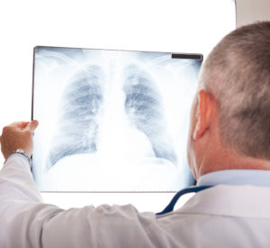
A chest X-ray procedure is an invaluable diagnostic tool, and something that everyone should understand a little better, especially if you have COPD.
What A Chest X-ray Can Tell You
The purpose of a chest X-ray is to see a flat image of your lungs, heart, airways, and the bones in your chest and spine. If a person experiences symptoms like shortness of breath or chest pains, a chest X-ray is usually the first diagnostic tool used.
By reading a chest x-ray, your doctor can determine if your symptoms are the result of a heart issue, or if you have a collapsed lung, pneumonia, a broken rib, emphysema, cancer, or other related health condition.
Once a particular diagnosis is made, there may be a series of chest X-rays to assess the progress of the disease in a less invasive way than other tests. Additional tests like a CT scan may also be performed to acquire a 3D picture rather than a flat image. These other tests will produce a more complex and detailed image compared to the chest X-ray.
COPD and Chest X-rays
Common complaints and symptoms of Chronic Obstructive Pulmonary Disease (COPD) include coughing and wheezing, an excess of mucus, a tightness in the chest, and difficulty breathing or catching one’s breath.
A chest X-ray will show significant signs of COPD with the following abnormalities:
- Lungs become enlarged, which is termed as hyperinflation. This occurs when the lung becomes damaged and loses its elasticity.
- Certain structural changes often occur with COPD. In this case, the lungs push up against the diaphragm, causing it to become flattened.
- Another obvious change is the shape of the heart, which will become narrowed or appear elongated in shape due to the inflated lungs.
- Bullae or pockets of air begin to appear within the lungs. Emphysema damages the lung tissue and can become progressively worse over time.
Final Diagnosis
In conjunction with a chest X-ray, other tests will most certainly be performed to arrive at a definitive diagnosis of COPD. From there, the doctor can determine the staging of the disease and recommend a treatment plan.
Contact Southwest Diagnostic Imaging Center if you experience shortness of breath or have excess mucus with coughing, as these are both prominent warning signs of COPD.