
MRI Exams in Dallas, TX
Providing a Comprehensive Range of Cutting-Edge MRI Services to Dallas
When it comes to detailed imaging for more accurate and precise diagnoses, an MRI is a great device. Southwest Diagnostic Imaging Center, features the latest in MRI equipment and is one of the most progressive freestanding outpatient imaging centers in Dallas since 1985. If you’re in or near the Dallas or Forth Worth area, call our diagnostic imaging center at (214) 345-6905 request your appointment online.
Musculoskeletal Imaging
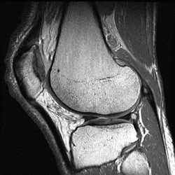 MRI is often used to study the knee, ankle, foot, shoulder, elbow, wrist, and hand. MRI is also a highly accurate method for evaluating soft tissue structures, such as tendons and ligaments, which can be seen in great detail with this method. Many subtle injuries are easily detected. In addition, MRI is used for the diagnosis of spinal problems, including disc herniation, spinal stenosis, and spinal tumors.
Musculoskeletal Imaging is an area of radiology that provides diagnostic imaging examinations of the skeleton, ligaments, muscles and joints. Specifically, musculoskeletal imaging is used to diagnose conditions that include arthritis, sports injuries, trauma and tumors. Procedures vary from general x-ray examinations to more complex ones such as joint aspiration, arthrography, biopsies, and stereotactic injections.
MRI examinations (magnetic resonance imaging or scanning) are used in musculoskeletal examinations because of their particular value and usefulness in imaging and evaluating soft tissue structures such as cartilage, muscles, bone marrow, nerves and vascular structures.
MRI Arthrography
MRI Arthrography is a two-step examination, an arthrogram, and an MRI exam. It is performed because of its exceptional diagnostic accuracy when there is a need to examine large joints like the shoulder, knee, and hip and small joints such as the ankle, elbow, and wrist. It is also used for complex cases such as postoperative knee and postoperative shoulder evaluations. Prior to the MRI exam, an arthrogram is performed in which a contrast agent (dye) is injected into a joint using x-ray fluoroscopy for guidance. The dye allows better visualization of the joint and possible tears of tendons or cartilage on the images produced by the MRI exam.
After the dye has been absorbed into the joint, the MRI exam is performed. The MRI generates different views of the joint and together with the dye, produces highly detailed images with extremely accurate information for diagnosis or evaluation.
Imaging of the Heart
MRI of the heart, aorta, coronary arteries and blood vessels is a tool for diagnosing coronary artery disease and other heart problems. Using MRI, our Dallas radiologists can examine the size and thickness of the chambers of the heart and determine the extent of damage caused by a heart attack or heart disease.
Some of the common uses of a heart MRI include:
- Evaluate symptoms of coronary artery disease, such as blood flow to the heart
- Evaluate scarring caused from a heart attack
- Diagnosing inflammatory conditions of the heart
- Diagnosing cardiovascular tumors
- Diagnosing cardiovascular infections
- Evaluating the anatomy of the heart chambers, valves and blood flow through vessels.
- Planning treatment for heart disease
Imaging of the Head and Spine
 Neuroradiology is a sub-specialty of radiology that specializes in performing diagnostic imaging procedures to diagnose and treat diseases and conditions of the central and peripheral nervous system in the brain, spine, head and neck. Neuroradiologists utilize a variety of imaging modalities such as CT and MRI imaging to obtain accurate and detailed information essential for interventional and surgical procedures.
MRI for neurological/brain imaging and spine studies provides outstanding image quality for diagnosis. The MRI software offers many mode and viewing options including the ability to reconstruct and rotate images to show soft tissue of the brain. MRA studies offer enhanced images of vascular structures.
Imaging for Cancer and Functional Disorders
Organs of the chest and abdomen, such as the liver, lungs, kidney and other abdominal organs, can be examined in great detail with MRI. This aids in the diagnosis and evaluation of tumors and functional disorders. For early diagnosis of breast cancer, MRI is a supplement to traditional mammography. Furthermore, because no radiation exposure is involved, MRI is often used for examination of the male and female reproductive systems.
Imaging for Prostate Disorders
A prostate MRI uses radiofrequency to create detailed images of the prostate gland and surrounding tissues.
Like CT scans, MRI scans show detailed images of soft tissues in the body. But MRI scans use radio waves and strong magnets instead of x-rays. A contrast material called gadolinium may be injected into a vein before the scan to better see details.
MRI scans can give a very clear picture of the prostate and show if the cancer has spread outside the prostate into the seminal vesicles or other nearby structures. This can be very important in determining your treatment options. But like CT scans, MRI scans aren’t usually needed for newly diagnosed prostate cancers that are likely to be confined to the prostate based on other factors.
Schedule Your MRI at Southwest Diagnostic Imaging Center in Dallas
Open 7 days a week, Southwest Diagnostic Imaging Center is a full-range, experienced, state-of-the-art imaging center in Dallas. Our mission is to provide unsurpassed patient care, the most advanced diagnostic imaging technology, and the highest standards of quality. If you are in need of a MRI, please contact our office at (214) 345-6905 to schedule an appointment today!